Root Surface Area of Permanent Mandibular Teeth in Patients with Anterior Open Bite Malocclusion: A CBCT Assessment
DOI:
https://doi.org/10.14456/nujst.2023.29Keywords:
Root surface area, Anterior open bite, Cone-beam computed tomography, Occlusal hypofunction, Orthodontic treatmentAbstract
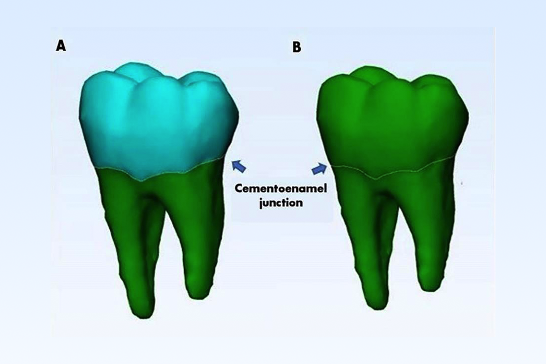
A previous study reported that the root surface areas (RSA) of the maxillary central and lateral incisors were substantially smaller in patients with anterior open bite malocclusion. However, the RSA of permanent mandibular teeth in patients with anterior open bite malocclusion has never been explored. The objective of this study was to investigate the RSA of permanent mandibular teeth in patients with anterior open bite malocclusion. Cone-beam computed tomography (CBCT) images of permanent mandibular teeth were selected from sixteen patients with anterior open bite malocclusion and sixteen patients with an anterior normal overbite. Mimics research software was used to construct three-dimensional tooth models from the CBCT images. 3-Matic Research software was employed to calculate the RSA. An independent t-test (p < 0.05) was used to compare the RSA of each tooth type. From the permanent mandibular central incisor toward the permanent mandibular second premolar, the means of RSA were significantly lower in the anterior open bite group than in the anterior normal overbite group. Anterior open bite malocclusion may affect the RSA in all permanent mandibular teeth except in permanent mandibular molars.
References
Arriola-Guillén, L. E., Valera-Montoya, I. S., Rodríguez-Cárdenas, Y. A., Ruíz-Mora, G. A., Castillo, A. A.-D., & Janson, G. (2020). Incisor root length in individuals with and without anterior open bite: a comparative CBCT study. Dental Press Journal of Orthodontics, 25, 23e21-23e27. https://doi.org/10.1590/2177-6709.25.4.23.e1-7.onl
Artese, A., Drummond, S., Nascimento, J. M. d., & Artese, F. (2011). Criteria for diagnosing and treating anterior open bite with stability. Dental Press Journal of Orthodontics, 16, 136-161. https://doi.org/10.1590/S2176-94512011000300016
Chakraborty, P., Chandra, P., Tandon, R., Azam, A., & Rastogi, R. (2020). Evaluation of Tongue Force on Mandibular Incisor in Various Malocclusions. Journal of Oral & Dental Health, 4, 7-11. http://doi.org/10.33140/jodh.04.02.02
Gu, Y., Tang, Y., Zhu, Q., & Feng, X. (2016). Measurement of root surface area of permanent teeth with root variations in a Chinese population—A micro-CT analysis. Archives of Oral Biology, 63, 75-81. https://doi.org/10.1016/j.archoralbio.2015.12.001
Harris, E. F., & Butler, M. L. (1992). Patterns of incisor root resorption before and after orthodontic correction in cases with anterior open bites. American Journal of Orthodontics and Dentofacial Orthopedics, 101, 112-119. https://doi.org/10.1016/0889-5406(92)70002-R
Hayashi, A., Hayashi, H., & Kawata, T. (2016). Prevention of root resorption in hypofunctional teeth by occlusal function recovery. The Angle Orthodontist, 86, 214-220. https://doi.org/10.2319/012215-47.1
Hujoel, P. P. (1994). A meta-analysis of normal ranges for root surface areas of the permanent dentition. Journal of clinical periodontology, 21, 225-229. https://doi.org/10.1111/j.1600-051X.1994.tb00310.x
Kaneko, S., Ohashi, K., Soma, K., & Yanagishita, M. (2001). Occlusal hypofunction causes changes of proteoglycan content in the rat periodontal ligament. Journal of periodontal research, 36, 9-17. https://doi.org/10.1034/j.1600-0765.2001.00607.x
Kuharattanachai, K., Jotikasthira, D., Sirabanchongkran, S., Srisuwan, T., Rangsri, W., & Tripuwabhrut, K. (2022). Three-dimensional volumetric evaluation of dental pulp cavity/tooth ratio in anterior open bite malocclusion using cone beam computed tomography. Clinical Oral Investigations, 26, 1997-2004. http://doi.org/10.1007/s00784-021-04179-x
Kuharattanachai, K., Rangsri, W., Jotikasthira, D., Khemaleelakul, W., & Tripuwabhrut, K. (2022). Does pulp cavity affect the center of resistance in three-dimensional tooth model? A finite element method study. Clinical Oral Investigations, 26, 6177-6186. http://doi.org/10.1007/s00784-022-04567-x
Lin, L.-H., Huang, G.-W., & Chen, C.-S. (2013). Etiology and Treatment Modalities of Anterior Open Bite Malocclusion. Journal of Experimental & Clinical Medicine, 5, 1-4. https://doi.org/10.1016/j.jecm.2013.01.004
Mamasoliyevna, D. S. (2023). Modern aspects of etiology and pathogenesis periodontal diseases. International Scientific Research Journal, 4, 141-151. https://doi.org/10.17605/OSF.IO/DXW8K
Mizrahi, E. (1978). A Review of Anterior Open Bite. British Journal of Orthodontics, 5, 21-27. https://doi.org/10.1179/bjo.5.1.21
Motokawa, M., Terao, A., Karadeniz, E. I., Kaku, M., Kawata, T., Matsuda, Y., . . . Tanne, K. (2013). Effects of long-term occlusal hypofunction and its recovery on the morphogenesis of molar roots and the periodontium in rats. The Angle Orthodontist, 83, 597-604. https://doi.org/10.2319/081812-661.1
Mowry, J. K., Ching, M. G., Orjansen, M. D., Cobb, C. M., Friesen, L. R., MacNeill, S. R., & Rapley, W. (2002). Root Surface Area of the Mandibular Cuspid and Bicuspids. Journal of Periodontology, 73, 1095-1100. https://doi.org/10.1902/jop.2002.73.10.1095
Ng, C. S. T., Wong, W. K. R., & Hagg, U. (2008). Orthodontic treatment of anterior open bite. International Journal of Paediatric Dentistry, 18, 78-83. https://doi.org/10.1111/j.1365-263X.2007.00877.x
Pan, J.-H., Chen, S.-K., Lin, C.-H., Leu, L.-C., Chen, C.-M., & Jeng, J.-Y. (2004). Estimation of single-root surface area from true thickness data and from thickness derived from digital dental radiography. Dentomaxillofacial Radiology, 33, 312-317. https://doi.org/10.1259/dmfr/19746488
Priya, S. P., Higuchi, A., Fanas, S. A., Ling, M. P., Neela, V. K., Sunil, P. M., . . . Kumar, S. (2015). Odontogenic epithelial stem cells: Hidden sources. Laboratory Investigation, 95, 1344-1352. https://doi.org/10.1038/labinvest.2015.108
Sang, Y.-H., Hu, H.-C., Lu, S.-H., Wu, Y.-W., Li, W.-R., & Tang, Z.-H. (2016). Accuracy Assessment of Three-dimensional Surface Reconstructions of In vivo Teeth from Cone-beam Computed Tomography. Chinese Medical Journal, 129, 1464-1470. http://doi.org/10.4103/0366-6999.183430
Selvi, A. K., Shanmugham, K. G., & Kannan, M. S. (2019). A Review on Role of Tongue in Malocclusion. Indian Journal of Public Health Research & Development, 10, 1067-1074. https://doi.org/10.37506/v10%2Fi12%2F2019%2Fijphrd%2F192272
Shetty, S. K., Ameena, B., Y, M. K., & Madhur, V. K. (2021). Root Resorption with Tads. Scholars Journal of Dental Sciences, 7, 247-251. https://doi.org/10.36347/sjds.2021.v08i07.012
Sringkarnboriboon, S., Matsumoto, Y., & Soma, K. (2003). Root Resorption Related to Hypofunctional Periodontium in Experimental Tooth Movement. Journal of Dental Research, 82, 486-490. https://doi.org/10.1177/154405910308200616
Studen-Pavlovich, D., & Vieira, A. M. (2019). The Dynamics of Change. In A. J. Nowak (Ed.), Pediatric Dentistry: Infancy through Adolescence (6th ed., pp. 555-561). https://doi.org/10.1016/B978-0-323-60826-8.00037-7
Suteerapongpun, P., Sirabanchongkran, S., Wattanachai, T., Sriwilas, P., & Jotikasthira, D. (2017). Root surface areas of maxillary permanent teeth in anterior normal overbite and anterior open bite assessed using cone-beam computed tomography. Imaging Science in Dentistry, 47, 241-246. https://doi.org/10.5624/isd.2017.47.4.241
Suteerapongpun, P., Sirabanchongkran, S., Wattanachai, T., Sriwilas, P., & Jotikasthira, D. (2018). Root Surface Areas of Maxillary Permanent Teeth in a Group of Thai Patients Exhibiting Anterior Open Bite Using Cone-Beam Computed Tomography. Chiang Mai Dental Journal, 39, 69-76. Retrieved from https://he01.tci-thaijo.org/index.php/cmdj/article/view/195876
Tasanapanont, J., Apisariyakul, J., Tanapan Wattanachaii, T., Sriwilas, P., Midtbø, M., & Jotikasthira, D. (2017). Comparison of 2 root surface area measurement methods: 3-dimensional laser scanning and cone-beam computed tomography. Imaging Science in Dentistry, 47, 117-122. https://doi.org/10.5624/isd.2017.47.2.117
Uehara, S., Maeda, A., Tomonari, H., & Miyawaki, S. (2013). Relationships between the root-crown ratio and the loss of occlusal contact and high mandibular plane angle in patients with open bite. The Angle Orthodontist, 83, 36-42. https://doi.org/10.2319/042412-341.1
Uribe, F. A., Janakiraman, N., & Nanda, R. (2015). Esthetics and biomechanics in orthodontics. In. R. Nanda (Series Ed.), Management of Open-Bite Malocclusion (2nd ed., pp. 147-179). https://doi.org/10.1016/B978-1-4557-5085-6.00009-6
Yun, H.-J., Jeong, J.-S., Pang, N.-S., Kwon, I.-K., & Jung, B.-Y. (2014). Radiographic assessment of clinical root-crown ratios of permanent teeth in a healthy Korean population. The journal of advanced prosthodontics, 6, 171-176. https://doi.org/10.4047/jap.2014.6.3.171

Downloads
Published
How to Cite
Issue
Section
License
Copyright (c) 2023 Naresuan University Journal: Science and Technology (NUJST)

This work is licensed under a Creative Commons Attribution-NonCommercial 4.0 International License.


